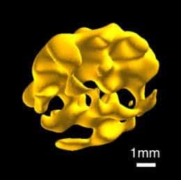
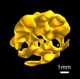
Researchers at Yale University have successfully mange to utilize a novel MRI technique to 3-D image the insides of hard and soft solids, like bone and tissue, opening the way for a new array of applications, like previously difficult to image dense objects.

The Yale scientists have developed a new method for MRI imaging, which they call “quadratic echo MRI of solids,” that works by targeting phosphorus atoms instead of hydrogen atoms. A more complicated sequence of radio waves pulses are fired for them to interact with phosphorus, a fairly abundant element in many biological samples, allowing for high-spatial-resolution imaging.
In the paper published recently in the journal PNAS, the Yale team report on various experiments designed to generated 3D MRIs using the phosphorus technique. They thus performed high-resolution 3D images of ex vivo animal bone and soft tissue samples, including cow bone and mouse liver, heart, and brains.
“This study represents a critical advance because it describes a way to ‘see’ phosphorus in bone with sufficient resolution to compliment what we can determine about bone structure using x-rays,” said Insogna, a professor at Yale School of Medicine and director of the Yale Bone Center. “It opens up an entirely new approach to assessing bone quality.”
The researchers say this new type of MRI would complement traditional MRI, not supplant it. MRI of solids should also be possible with elements other than phosphorus, they say.
The researchers believe this new type of MRI imaging should be used to complement the traditional MRI already in place, and claim that MRI imaging of solids through other elements other than hydrogen or phosphorus should be possible. The quadratic echo MRI technique, however, can’t be used on living beings – for one it generates way too much heat. Immediate applications include archaeology, geology, oil drilling.


